Shannon Clinic – Melbourne Chiropractic and Sports Care Blog
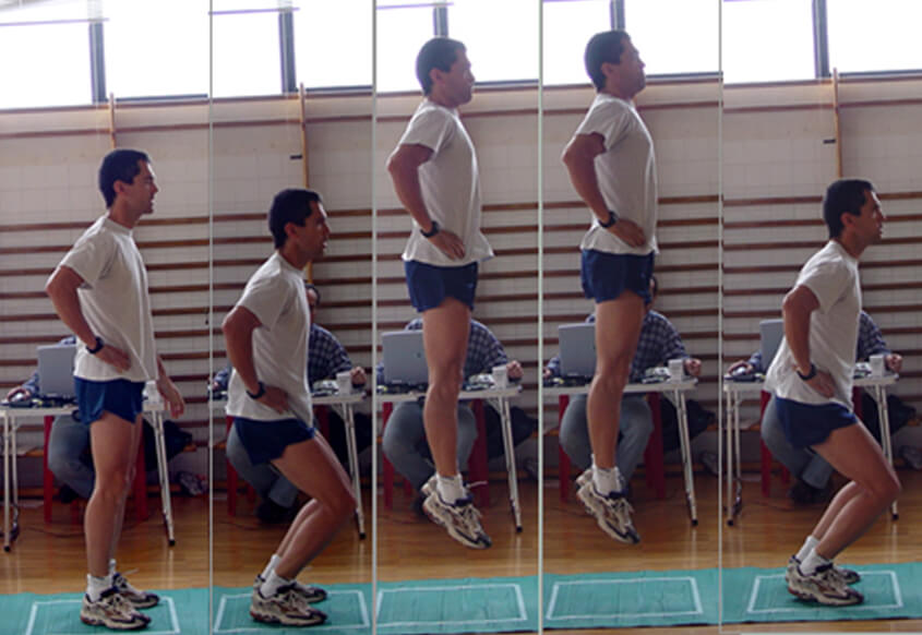
Running related injuries (RRI) have remained reasonable unchanged since the early 1980s and range from 17-79%. RRI are the most prevalent reason why runners cease participation with the most commonly affected areas being the knee and lower leg. Studies have looked at demographic and anthropometric factors to determine risk factors for RRI injuries with consideration given to age, sex, BMI. These factors were found to be associated with certain injuries where females are more likely to sustain anterior knee and ITB pain, while men were more likely to suffer from Achilles tendinopathy and plantar fasciitis.
A recent study by Hollander, et.al anchored by expert running biomechanists Irene Davis, retrospectively looked at 550 recreational runners to determine the different factors that might play a role in RRI and to give further weight to the notion that RRI are multifactorial. Their study examined biomechanics, demographics and anthropometric factors.
Running Related Injury Risks
Their paper found the foot striking patterns were associated with certain injuries; Achilles injuries were 2 times more likely in those with a midfoot strike pattern. This is potentially a result of the changes in Achilles loading due to the position of the foot and ankle on impact. Posterior leg injuries (most commonly calf injuries) were associated with forefoot strike patterns.
They also found that higher peak vertical ground reaction forces, the forces directly impacted on the body as the foot hits the ground were associated with hip and groin pain. Interestingly though, they didn’t find an association between cadence (steps per minute) and injury location, whereas other studies have found that a lower cadence is associated with anterior (front) leg pain.
Key Injury Risk Factors
Some of the key overall injury risk factors Hollander found indicate your risk increases for:
An Achilles injury if you are older, male and a midfoot striker
An ITB injury if you are older
A hip/groin injury if you are female
A thigh injury or anterior knee pain if you are female
A patella or quadriceps tendinopathy if you are male.
How To Avoid A Running Injury
The most important factor to mitigate the risk of a RRI is appropriate load management. This means periodizing training, having appropriate rest periods and deload weeks of training, and to scale up training in roughly 10% increments each week.
Tendinopathies are extremely prevalent in running including Achilles, plantar fasciitis, glute medius, patellar and quadriceps. Tendinopathies are directly associated with load levels however, the best way to prevent tendinopathies is by having strong tendons. Therefore a gym based strengthening program is vitally important for runners. There is also evidence to support the use of minimal footwear to improve intrinsic foot muscle strength with a 2018 paper showing over a 12 week period, minimal shoes were as effective at improving intrinsic foot muscle strength as performing a foot strengthening program.
Identifying and correcting any technique and/or biomechanical deficiencies will ensure a more evenly loaded musculoskeletal system in addition to reducing high peak vertical growth reaction forces both of which are related to RRI.
Finally, make sure you rehabilitate any injuries you have sustained as prior injuries, especially soft tissue injuries such as calf strains are a risk factor for a subsequent injury. Anecdotally, in our Melbourne city chiropractic clinic the three most prevalent factors leading to injury are poor training load management, muscle imbalances especially around the pelvis and poor technique. To find out how our sports chiropractic clinic manages training loads click here. Additionally, if you are interested to learn how the weight of your running shoes might be slowing you do, you can read more here. For those keen to explore the benefits of minimal shoes we prefer Sole Mechanic Footwear, who specialise in minimal shoes. If you use the code “SHANNONCLINIC7” you will receive a 15% discount.
If you are experiencing a running related injury or encounter repeated running related injuries, Having worked with state and national track and field athletes, as well as endurance and ultra endurance athletes Melbourne city chiropractoc Dr. Shannon is well placed to assess your running related injury. You can make an appointment below. Our Melbourne chiropractic clinic is conveniently located in the heart of the Melbourne CBD on Collins Street in the Manchester Unity building, opposite the Melbourne Town Hall and City Square.
Shannon Clinic – Melbourne Chiropractic and Sports Care Blog
It is with great pleasure that melbourne city sports chiropractor can share his paper,“Common and Less Well-Known Upper Limb Injuries in Elite Tennis Players”led by Dr. Shannon and co-authored by my esteemed colleagues Dr. Timothy Wood, Dr. John Kelly IV and Dr. Brian Cable in the October 2020 issue of the American College of Sports Medicine’s Current Sports Medicine Reports journal, with great thanks going to Dr. Matthew Gammons.
Key Upper Limb and Injury Findings In Elite Tennis Players
Nicole Melichar Martinex and Demi Schuurs practice at the Australian Open
Our paper is the first of its kind to review injury data in solely elite male and female players drawn from the most recent epidemiological studies including data from the US Open, Australian Open, Wimbledon, Davis Cup, ATP and WTA. Our paper highlights the common injuries seen in the upper limb of elite players but additionally highlights less well-known injuries that occur in the upper limb which are not routinely reported on in the literature. These injuries include posterior shoulder instability, medial elbow ulnar collateral injuries, distal humeral bone stress injuries, nondominant wrist ulnar ligament injuries. Furthermore, we highlight the discrepancies that still exist in how injuries are recorded in elite tennis, even in light of a consensus statement recommendation, as well as the importance of player workloads and the relationship with injury risk.
We note that muscle strain and tendon injuries are the most prevalent injuries seen in elite players with higher rates of chronic injuries in the upper limb. However, there is an argument to say that many of the acute injuries that occur may well be due to suboptimal rehabilitation of a long-standing injury and/or returning to play too early. A paper published in 2020 applied the acute:chronic workload ratio to junior tennis players which found that players with an acute:chronic workload ratio greater than 1.5 in the previous week sustained an injury. From Melbourne city sports chiropractor Dr. Shannon’s experience working with elite professional and junior players these findings appear to hold true. To find out how Dr. Shannon manages athletes training load you can read more here.
Further Research into Tennis Workloads
Subsequentially, a key area of further research is establishing safe workloads for elite tennis players to assist in minimizing injury risk. A part of this process may well involve a supervised injury prevention program specific to the needs of the elite player, those of which have been successful in other sports such as soccer. Notwithstanding, managing playing loads of the elite tennis player are an ongoing challenge with quick turnaround times between matches, varied lengths of matches, the number of matches played in one day (singles, doubles, mixed doubles in grand slams) and across a tournament, the long season and short off season, in addition to the financial pressures to keep playing to stay on tour and improve one’s ranking. This challenge is routinely reminded to us and was particularly evident in 2017/18 where there were exceptionally high numbers of injuries in top ranked players.
As clinicians, together with researchers and competition organizers it is imperative that we are able to work towards a competition where players are optimized, performing at their highest levels, rewarded equally (men and women) for their performance and where injury risks are mitigated through proper recovery and injury rehabilitation programs, making sure players are safe to return to play, as well as having time away from the court (a genuine off season). If you are interested in learning more about specific shoulder injuries, many of which are seen in tennis players, you can read the paper Melbourne city chiropractor Dr. Shannon’s co-authored with Dr. John Kelly IV on shoulder injuries here.
With a wealth of experience working with recreational, junior elite and tour professional players Melbourne city chiropractor Dr. Shannon is well placed to help assess and treat tennis based injuries. You will find our Melbourne CBD chiropractic clinic in the Manchester Unity building on the corners of Collins Street and Swanston Street, opposite the Melbourne Town Hall and City Square.
Shannon Clinic – Melbourne Chiropractic and Sports Care Blog
Anterior cruciate ligament (ACL) knee injuries are devastating injuries that can significantly impact an athlete’s career including ending their ability to return to high level competitive sport. Individuals who suffer an ACL injury have lower self-reported knee function, quality of life and a greater risk of long-term joint morbidity including early osteoarthritis (1,2). Whilst those who undergo reconstructive surgery have lower return to sport rates with relatively high reinjury rates up to 24%, with the greatest risk being in the first 7 months (1,3).
The mean time to return to sport following ACL reconstructive surgery (ACLR) is approximately 7 months with accelerated rehabilitation program as short as 6 months (4,5). However, it has been established that when athletes return to sport following an ACLR they continue to exhibit neuromuscular and biomechanical alterations including quadriceps strength deficits resulting in altered landing patterns (1,6). These deficits may potentially result in higher risks of reinjury to the grafted and/or contralateral knee, all indicating that potentially we are returning our athletes too quickly to play following ACLR.
This study examines professional soccer players specifically looking at kinetics, focusing on asymmetries during a counter movement jump (CMJ). The framework for this article is an analytical observational cross-sectional design and although it is not the most robust design format, it enables a cohort to be observed and compared at certain time points across the study. It executes this by dividing athletes into 4 groups, those who are 3-6 months, 6-9 months, >9 months into their ACLR rehabilitation and a control group. Strengths to this observational study are the use of the CMJ as an assessment measurement tool provided the risk of errors is minimized, as well as the use of a control group. Some questions are raised over the participation inclusion criteria as there is no mention of; any current or prior ankle injuries which are prevalent in soccer and can effect landing mechanics (7,8,9,10), which knee was injured dominant or non-dominant leg, if strength asymmetries were present prior to the injury, whether all ACLR participants followed a standardised rehabilitation program, were any players involved with ACL injury prevention programs and the cohort is specific to professional soccer players.
Counter Movement Jump
Findings
The study found that jump height (which is linked to an ability to complete tasks at a high level in soccer such as sprinting and change of direction) increased in the first 3-6 months however, it plateaued at 6-9 months and remained well below (3-4cm) the control group after 9 months. Whilst peak power, as measured by dual force plates followed a similar pattern and remained well below (3-4W/kg) the control group after 9 months. There were also significant interlimb asymmetries in the ACLR group during the eccentric (preload and deceleration phases) and concentric (jump phase) which decreased the further out from surgery the participants were, yet significant differences remained after 9 months. Indicating ACLR players were employing an offloading strategy to protect the injured knee. Furthermore, the uninjured limb was the dominant force producer. Both findings are consistent with other similar research indicating the changes occur due to altered nervous system function, strength deficits, reduced range of motion and fear of reinjury. The over reliance on the dominant limb for peak force production is important as it results in greater torque and stress loading of the knee and if the musculature is unable to dissipate the force effectively during a jump landing it may contribute to higher risk of injury, it may also lead to fatigue and potential injury of the uninjured knee.
Results
The results suggest that even after 9 months soccer players who have undergone ACLR are showing power, strength, and asymmetries between limbs with lower power and jump height figures compared to healthy controls. Knowing that strength deficits are associated with reinjury rates in a variety of lower limb injuries, it would suggest that potentially players are being returned to play before they are ready, increasing their risk of reinjury (11,12).
Although this study has its weaknesses the results are consistent with those in similar studies looking a landing patterns in participants following ACLR helping to build on the available evidence that limb kinetic asymmetries exist in ACLR patients up to and greater than 9 months (1,6,13). These findings together suggest that longer recovery times are warranted, and individuals should only be returned to play following ACLR when they have limb asymmetries within a tolerable limit. Read et.al recommend benchmark goals such as, a jump height of 33-35cm and a concentric impulse asymmetry of no more than 2.5-3.1%. They also provide quartile figures, enabling clinicians to establish whether an individual is progressing quickly or slowly with their rehabilitation based on the CMJ metrics. They also point out that inter limb differences are task, variable and physical quality specific, meaning limb differences will occur across different tests and variables in the same task therefore using one asymmetry metric such as a single leg hop test with a <10% asymmetry isn’t an appropriate guide to determine progression and return to play status. A combination of tasks should be used including single leg hop, isokinetic strength, CMJ analysing the different variables in the task ie. height, power etc.
Concluding Thoughts
ACL injuries are devastating knee injuries and it is paramount to reduce the risks of reinjury that athletes are not rushed back prematurely, furthermore evidence shows a 50% reduction in reinjury rates for every month return to sport is delayed up to 9 months post-surgery (5). Focusing on reducing lower limb strength asymmetry, especially improving quadriceps strength is vitally important in helping to monitor an athletes ACLR progress and in determining when they are ready to return safely to sport. Consideration should also be given to ACL injury prevention programs which have been shown to reduce the risk of ACL injures by 53%, with any program consisting of strength, plyometrics, agility, balance and flexibility exercises (2).
With evidence particularly in soccer showing a high rate of reinjuries it is important that athletes are not rushed back into their sport. Melbourne ciry chiropractor Dr. Shannon is well placed to help you navigate your injury assessment, rehabilitation and return to sport. To make an appointment to see sports chiropractor Dr. Shannon or remedial massage therapist Paula Pena click below. You will find our chiropractic clinic centrally located on Collins Street in the CBD of Melbourne.
Shannon Clinic – Melbourne Chiropractic and Sports Care Blog
In mid to late 2016, Melbourne city chiropractor Dr. Shannon co-authored a paper on shoulder injuries with colleague Dr John Kelly IV orthopaedic surgeon at the University of Pennsylvania in the American Journal of Family Medicine. It was a bitter sweet achievement, with the hard work that went into the paper being offset by the disappointing finding that the journal was a predatory journal. Upon finding this news out John and Melbourne sports chiropractor Dr. Shannon made the difficult choice to withdraw our paper from the journal. We are still very pleased to be able to share it here.
An Evidence Based Approach to Shoulder Injuries
Shannon N1* and Kelly IV JD2*
1Sports Chiropractor, 9/220 Collins Street, Melbourne, Victoria, Australia 2Director of Shoulder Sports Medicine, University of Pennsylvania, USA
Introduction
Shoulder pain is a complex and prevalent complaint for primary care physicians with up to 66.7% of the population experiencing shoulder pain at some point in their lifetime [1]. It is therefore important that primary care physicians have a thorough understanding into the anatomical and kinematic make up of the shoulder joint.
The glenohumeral joint is formed by the humeral head and the glenoid fossa of the scapula [2]. This creates a ball and socket joint, which results in a joint with a remarkable range of motion, but the trade off, is a joint which has a loss of biomechanical stability [2]. This is due to a humeral head with a large spherical shape articulating with a small fossa, similar to a golf ball and tee [2]. The stability of the joint comes from the surrounding soft tissue structures which includes the articular cartilage (labrum), the joint capsule and ligaments (coracohumeral ligament, superior, middle and inferior glenohumeral ligaments), the rotator cuff muscles (supraspinatus, infraspinatus, subscapularis, teres minor) which supply the concave joint compression [2].
Movement at the glenohumeral joint is dependent on three other joints, the scapulothoracic, acromioclavicular and sternoclavicular joints. Together the movement that occurs between these 4 joints is called “scapulohumeral rhythm” [3]. During normal movement of the shoulder the scapula will upwardly and posteriorly tilt on the thorax during elevation of the arm in flexion, abduction, scapular plane abduction or unrestricted overhead reaching [4]. Scapulothoracic internal and external rotation are more inconsistent and are determined by the plane in which the arm is being elevated in and on what portion of elevation range of motion is considered. The motion of the scapula is dependent on scapular rotating muscular balance, with excessive internal rotation, or protraction, the result of muscle weakness. The scapular must adjust in the transverse plane for the intended plane of elevation [5].
The complexity of the joint complex, coupled with the lack of biomechanical stability of the glenohumeral joint exposes the joint to injury [6]. The more common mechanism of injuries to the shoulder include direct and indirect trauma to the shoulder (FOOSH injury, direct blow to the shoulder, high force impact to the shoulder, lifting a heavy object), repetitive overuse and disruption to the scapulohumeral rhythm [3,5-8]. From here injuries can be classified as acute, chronic, stable, unstable, strains, tears (partial thickness, full thickness, degenerative). It is also important to consider the cervical and thoracic spine when evaluating a suspected shoulder injury as there can be concomitant injury to both [9].
Exercise also plays an important role in determining what type of shoulder injury has been sustained, with different injuries associated with different sports. Table 1 provides a list of sports, the common types of shoulder injuries seen in that sport, and the more common mechanism of injuries.
There is an exhaustive list of potential injuries that can occur with the shoulder. This manuscript will provide an evidence based overview on the more common shoulder injuries seen in clinical practice, how they should be evaluated, and what treatment options are available to primary care physicians.
Impingement
Shoulder impingement is a very common shoulder complaint which is believed to be associated with rotator cuff disease [23]. It can be broken down into different categories. There is external impingement, internal impingement, which can then be further divided into anterior, posterior and coracoid impingement [5].
External Impingement
External shoulder impingement is usually confined to the subacromial space and is associated with overhead overuse. The pain is commonly anterior with overhead activities, and is due to the compression or abrasion of the cuff tendons or long head of the biceps tendon beneath any aspect of the coracoacromial arch [24]. Common causes are structural and degenerative, such as acromion spurs, shape of the acromion, degenerated cuff tendon [25]. In patients under 40 years of age where external impingement is suspected, further investigation is required, as often there is will other factors involved such as instability, scapular dyskinesis. A protracted scapula will lessen the acromial humeral distance and potentiate impingement. Many patients can be success can be successfully treated with scapula rehabilitation instead of acromioplasty.
Internal Impingement and Posterior Capsular Tightness
Internal shoulder impingement usually occurs in younger athletes involved with overhead sports such as baseball, swimming, tennis. The pain is usually posterior and occurs in the late cocking phase of throwing. It is due to the humeral head loss of centricity in the glenoid fossa, either as a result of laxity in the anterior capsule, tightness in the posterior capsule, a type II labral injury, or scapular dyskinesis [25]. Internal impingement occurs when the posterior edge of the supraspinatus and the anterior edge of the infraspinatus impinge against the posterior superior glenoid and labrum in the late cocking position [26].
Another source of shoulder pain is due to microtrauma of the posterior shoulder capsule and cuff, leading to scarring and contracture causing a reduction in internal shoulder rotation ie. glenohumeral internal rotation deficit (GIRD) [25]. This then causes the humeral head to rotate posterior superiorly, impinging the rotator cuff between the humeral head and glenoid [25].
Assessment
Assessment should always consist of a thorough history and physical exam. The physical exam should look to confirm the impingement syndrome as well as the potential cause of the impingement including tendon pathology, scapular dyskinesis, capsule laxity or instability. Physical exam tests to assess for impingement include active and passive range of motion, Neer impingement sign, Hawkins test, horizontal adduction, resisted abduction, scapular assistance test and scapular retraction test [27-28]. Throwers should undergo a full kinetic chain evaluation, with particular attention paid to abduction weakness, lead and stance leg hip rotation deficits and quadriceps tightness.
If warranted, further imaging will be driven by the diagnosis and should include plain films [29]. Ultrasound and MRI are equally as good at picking up tendon pathology [29]. MRI and MRA (gold standard) are best for identifying labral tears [30].
Treatment
Scapular motion is a key factor when assessing and treating impingement with scapular motion abnormalities identified in subjects with impingement and rotator cuff disease [31]. Without correction of the dysfunctional shoulder kinematics the impingement mechanism will remain. Therefore a targeted stretching and strengthening program is important to assist in reducing pain and improving function [32]. It is important though to combine the exercise therapy with other treatments, as exercise alone is not as effective as combined treatments with exercise [33]. With combined therapies and treatments yielding better results than single interventions alone [33].
Acute patients tend to respond well to ultrasound guided cortisone injection, followed by a structured rehabilitation program, whereas those who have injections alone tended to fare worse [33,34]. Exercise therapy with specific exercises combined with kinesio tape and acupuncture are ideal for patients with early shoulder impingement. While low level light therapy and localised NSAID injections are not recommended [33]. Nitroglycerine patches may help cuff pain. PRP injections are currently inconclusive. Correction of kinetic chain abnormalities may require abductor strengthening, quadriceps and hip stretching as well as scapular repositioning exercises.
Surgical treatment options such as arthroscopic acromioplasty, bursectomy, subacromial decompressive surgery, labral and cuff repairs should be considered to aid in restoring normal function to an impinged houlder where the impingement is due to acromion spurring or glenohumeral joint instability or in long standing cases [33].
Labral Tears
The labrum is a dense fibrous tissue that is attached to the periphery of the glenoid fossa, increasing the depth and width of the glenoid fossa [35]. This results in greater stability of the shoulder while still allowing for increased range of motion. The labrum also serves as the attachment site for the long head of the biceps with 40 to 60% of the fibres inserting into the supraglenoid tubercle and the remaining into the labrum [36]. Other structures serving as attachment sites for the labrum include the long head of the triceps and glenohumeral ligaments.
Labral tears can occur at any point around the labrum, but the more common labral tears include SLAP tears (Superior Labral Anterior Posterior) and Bankart lesions [35].
SLAP Tears
The most common mechanisms of injury for a SLAP tear are forced traction on the shoulder such as a external rotation and hyperextension force, and direct compression (acute traumatic or chronic-overhead throwing athlete) [35]. There are four classes of SLAP tears [36]:
Type I: degenerative tear of the under surface of the superior labrum with the biceps anchor intact. Type II: tear of the superior labrum as well as of the biceps anchor. Type III: bucket-handle tear of the superior labrum with biceps anchor intact. Type IV: bucket-handle tear of the superior labrum with extension into biceps tendon
SLAP tears tend to present with deep pain in the shoulder which can be sharp or dull [35]. The pain can be aggravated by overhead activity, pushing or lifting a heavy object. There maybe catching, popping or clicking and often there will be other associated shoulder conditions present [35]. Throwing athletes may present with pain in the late cocking phase or early acceleration phase of throwing, which may also be accompanied by weakness or decreased velocity [25,35].
Bankart Lesions
Bankart lesions occur when the humeral head shifts anterior and inferior out of the glenoid fossa. In doing so it damages the anterior inferior labrum, glenohumeral ligaments, joint capsule and rotator cuff [35]. They are seen in 85-97% of anterior shoulder dislocations [37,38]. The end result is anterior instability as a result of the damaged structures, as well as the loss of negative pressure the labrum creates to hold the humeral head in the socket [35]. The main symptom of a Bankart lesion therefore will be instability and subsequent pain which is usually associated with a dislocation event or recurrent episodes of instability.
The key physical examination tests to evaluate for a labral tear or Bankart lesion include, Shift and Load, Andrews “pitcher” provocation test, Kim 1 and Kim 2, Sulcus sign, abduction with inferior distraction, O’Brien’s, Apprehension test, Crank test [28,35,39]. Kuhn’s Test comprises of Sulcus sign, abduction and inferior distraction and Yergason’s test. If all three are negative there is a 6% of a SLAP tear, if one is positive there is a 14-20% chance of a SLAP tear and if 2 or more are positive there is a 34-66% of a SLAP tear, which warrants an MRA [40]. MRI and MRA (gold standard) are best for identifying labral tears [30].
Treatment
The treatment of first time dislocated shoulders remains very controversial; however in the absence of significant glenoid or humeral bone loss, rehabilitation may be a viable option. The presence of a bony bankart lesion or large Hill Sachs lesion will likely be best treated with surgical repair.
Most surgical cases can be successfully managed with an arthroscopic approach. However, if glenoid bone loss exceeds 25%, then a coracoid transfer procedure (Latarjet) may be the best recourse. The initial treatment of SLAP labral tears should involve a period of conservative treatment for 6-12 weeks, which should include, rest, physical therapy and nonsteroidal anti inflammatories, with the goal of reducing pain and improving shoulder function [28]. Those failing conservative treatment and overhead throwing athlete’s, should consider arthroscopic repair which has shown predictably good functional results with an acceptable rate of return to play [36].
Rotator Cuff Tears
Rotator cuff pathology is associated with 30-70% of cases of shoulder pain with anywhere between 5-40% involving tears [41]. The rotator cuff muscles are comprised of the infraspinatus, supraspinatus, subscapularis and teres minor muscles. The cuff muscles together act to stabilize the humeral head in the glenoid fossa and to facilitate movement of the arm [42]. Injuries to the rotator cuff include strains, tendonitis (impingement syndrome) and tears.
There are two categories of tears, acute traumatic and chronic degenerative. Acute tears usually occur in younger athletes and often involve a FOOSH injury, high energy trauma, heavy lifting, shoulder dislocation [42]. Chronic degenerative tears occur in older individuals, with the incidents of rotator cuff tears increasing with age [43]. Many chronic tears tend to be asymptomatic and often involve chronic overuse, mechanical impingement, normal age degeneration of the tendon, and lack of blood supply to the tendon [42]. Additional risk factors include smoking, hypercholesterolemia and genetics [43].
Assessment
On examination there is usually pain on palpation, weakness with or without pain on manual muscle testing, pain when elevating the arm overhead and difficulty sleeping on the affected shoulder. There is no relationship between the severities of pain to the size of the tear [44]. But increased pain or worsening pain indicates tear progression. Orthopaedic assessment should include the Hawkins-Kennedy test, infraspinatus muscle test, and painful arc, as well as internal rotation lag sign, external rotation lag sign, lift off test, dropping sign as well as empty can and bear hug test [45].
Imaging
Imaging work up is useful in patients who present with 6 weeks or more of rotator cuff tears symptoms. Plain film X ray and ultrasound together are recommended as the most valid imaging method to exclude tendon rupture in those unresponsive to conservative management [41]. MRI is excellent at identifying full thickness tears. The evidence for the use of MRI to identify partial tears is conflicting [41,46]. MR arthrograms only have limited usefulness when compared with MRI [41]. There is no consensus statement yet as to which imaging method is more precise at identifying full and partial thickness tears.
Treatment
Conservative management over surgical repair for rotator cuff tears has been proven to be beneficial in some studies over the short and long term, while others have shown early surgical intervention provides better outcomes [46]. Conservative management needs to involve an individualised rehabilitation program that focuses on range of motion optimization, scapular stabilization and core abdominal strengthening. NSAIDs can be used in the first 3-4 weeks to manage pain levels, but in the long term they provide no benefit in restoring function to the shoulder [41]. In general, patients with two tendon tears are likely best served with surgical repair.
There is inconclusive evidence regarding the benefit of PRP injections in the treatment of rotator cuff pathology, although one recent study has shown PRP in association with surgical repair of medium to large tears improved the quality of the tendon, but not the healing rate [47].
Surgical management of rotator cuff tears
There is no obvious long term difference in open vs. arthroscopic cuff repair. However repairs performed with the arthroscopy do realize less pain. Furthermore, larger retracted tears are better visualized with arthroscopy so that mobilization and side to side repairs can be afforded. Subscapularis tears may be present in up to 35% of rotator cuff lesions and must be addressed in order to attain optimal results.
Clavicular Fracture
Clavicular fractures account for 35-45% of all shoulder girdle fractures. Middle ⅓ fractures account for 81% of all fractures, with 17% lateral ⅓ and 2% medial ⅓ [48]. The most common mechanismof injury involves a fall on the lateral shoulder which commonly occurs in cyclists and motorcycle pilots, less common causes include a direct blow or FOOSH injury [6].
It is uncommon to have secondary complications associated with the fracture, but pneumothorax, hemothorax and injuries to the brachial plexus and subclavian arteries have been reported [49]. Examination should therefore include a neurovascular and lung exam.
Plain film x ray is sufficient to identify the fracture with the Zanca, or cephalic view essential to assess displacement. Non displaced fractures in any region of the clavicle can be treated conservatively with a sling, ice and analgesics [6]. With elbow range of motion exercises started when tolerable. Shoulder range of motion and strengthening exercises should begin once the fracture has healed.
There is a slight increase in risk of delayed non-union in displaced fractures treated conservatively compared to surgical repair. Surgical repair is also associated with better function and less disability in the short term since clavicle strut shortening is associated with scapular protraction [48]. Displaced fractures may require surgical intervention if more than 2cm shortening is seen and scapula dyskinesis is present.
Conclusion
Shoulder pain is a complex, common complaint seen by primarycare physicians. It is pertinent that primary care physicians have a sounds understanding of the anatomy and kinematics of the shoulder, and understand the potential impact sports have on the shoulder. This knowledge coupled with an evidenced based approach to examination and management should enable primary care physicians to more accurately diagnose and more appropriately management common shoulder injuries.
If you made it this far congratulations and you might enjoy reading some of Melbourne city chiropractor Dr. Shannon’s other published works such as his article on injuries in elite tennis players and sports related concussion. If you would like to make appointment to see Melbourne CBD sports chiropractor Dr. Shannon or remedial massage therapist Paula Pena you can book online below. You will find our Melbourne city chiropractic clinic on Collins Street, opposite the Melbourne Town Hall.
Luime JJ, Koes BW, Hendriksen IJ, Burdorf A, Verhagen AP, Miedema HS, et al. Prevalence and incidence of shoulder pain in the general population; a systematic review. Scand J Rheumatol. 2004; 33: 73-81.
Rockwood CA, Matsen FA, Wirth MA, Lippitt SB, Fehringer EV, Sperling JW. The Shoulder, 4th ed., Elsevier Health Sciences. 2009; 12-25 p.
Brukner P and Kahn K. Clinical Sports Medicine 3rd ed., Mc Graw-Hill Professional. 2008; 244 p.
Mc Clure PW, Michener LA, Sennett BJ, Karduna AR. Direct 3-dimensional measurement of scapular kinematics during dynamic movement in vivo. J Shoulder Elbow Surg. 2001; 10: 269-277.
Ludewig PM and Braman JP. Shoulder Impingement: Biomechanical Considerations in Rehabilitation. Manual Ther. 2011; 16: 33-39.
Quillen MD, Wuchner M, Hatch RL. Acute Shoulder Injuries. Am Fam Physician. 2004; 70: 1947-1954.
Bey MJ, Elders GJ, Huston LJ, Kuhn JE, Blasier RB, Soslowsky LJ. The mechanism of creation of superior labrum, anterior and posterior lesions in a dynamic biomechanical model of the shoulder: The role of the inferior subluxation. J Shoulder Elbow Surg. 1998; 7: 397-401.
Baker CL, Uribe JW, Whitman C. Arthroscopic evaluation of acute initial anterior shoulder dislocations. Am J Sports Med. 1990; 18: 25-28.
Sembrano JN, Yson SC, Kanu OC, Braman JP, Santos ER, Harrison AK, et.al. Neck-shoulder crossover: how often to neck and shoulder pathology masquerade as each other? Am J Orthop. 2013; 42: 76-80.
Silberman MR. Bicycling Injuries. Curr Sports Med Rep. 2013; 12: 337-345.
Kelly BT, Barnes RP, Powell JW, Warren RF. Shoulder Injuries to Quarterbacks in the National Football League. Am J Sports Med. 2004; 32: 328-331.
Kuhn JE. Current Concepts: Rotator Cuff Pathology in Athletes – A Source of Pain or Adaptive Pathology. Curr Sports Med Rep. 2013; 12: 311-315.
Knesek M, Skendzel JG, Dines JS, Altchek DW, Allen AA, Bedi A. Diagnosis and Management of Superior Labral Anterior Posterior Tears in Throwing Athletes. Am J Sports Med. 2013; 41: 444-460.
Olson DE, Sikka RS, Hamilton A, Krohn A. Football Injuries: Current Concepts. Curr Sports Med Rep. 2011; 10: 290-298.
Wadsworth, TL. When Golf Hurts: Musculoskeletal Problems Common To Golfers. Curr Sports Med Rep. 2007; 6: 362-365.
Kim KS, Park JK, Lee J, Kang BY. Injuries in National Olympic Level Judo Athletes: an Epidemiological Study. Brit J Sport Med. 2015; 49: 1144-1150.
Roedl JB, Nevalainen M, Gonzalez FM, Dodson CC, Morrison W, Zoga AC. Frequency, Imaging Findings, Risk Factors, and Long-Term Sequalae of Distal Osteolysis in Young Patients. Skeletal Radiol. 2015; 44: 659-666.
Crichton J, Jones DR, Funk L. Mechanisms of Traumatic Shoulder Injury in Elite Rugby Players. Brit J Sport Med. 2012; 46: 538-542.
de Almeida MO, Hespanhol LC, Lopes AD. Prevalence of Musculoskeletal Pain Among Swimmers in an Elite National Tournament. Int J Sports Phys Ther. 2015; 10: 1026-1034.
Pieber K, Herceg M, Fialka C, Oberleitner G, Gruther W, Paternostro-Slug T. Is Suprascapular Neuropathy Common in High-Performance Beach Volleyball Players? A Retrospective Analysis. Wien Klin Wochenscher. 2014; 126: 655-658.
Reeser JC, Gregory A, Berg RL, Comstock, RD. A Comparison of Women’s Collegiate and Girls’ High School Volleyball Injury Data Collected Prospectively Over a 4 year Period. Sports Health. 2015; 7: 504-510.
Dines JS, Bedi A, Williams PN, Dodson CC, Ellenbecker TS, Altchek DW, et.al. Tennis Injuries: Epidemiology, Pathophysiology, and Treatment. J Am Acad Orthop Surg. 2015; 23: 181-189.
Michener LA, McClure PW, Karduna KR. Anatomical and Biomechanical mechanisms of subacromial impingement syndrome. Clin Biomech. 2003; 18: 369-379.
Scerpella TA. Chronic Injuries About The Adult Shoulder: Impingement,
Rotator Cuff Injuries and Instability. American College of Sports Medicine
Team Physician Course. February 2012. Lecture 8, San Antonio, (USA).
Paley KJ, Jobe FW, Pink MM, Kvitne RS, ElAttrache NS. Arthroscopic findings in the overhand throwing athlete: evidence for posterior internal impingement of the rotator cuff. Arthroscopy. 2000; 16: 35-40.
Silva L, Andreu JL, Munoz P, Pastrana M, Millan L, Sanz J, et.al. Accuracy of physical examination in subacrominal impingement syndrome. Rheumatology. 2008; 47: 679-683.
Nuri A, Evrim S, A Arya. Superior labrum anterior to posterior lesions of the shoulder: Diagnosis and arthroscopic management. World J Orthop. 2014; 5: 344-350.
Naqvi GA, Jadann M, Harrington P. Accuracy of ultrasonography and magnetic resonance imaging for detecting full thickness rotator cuff tears. Int J Shoulder Surg. 2009; 3: 94-97.
Ludewig PM, Reynolds JF. The association of scapular kinematics and glenohumeral joint pathologies. J Orthop Sports Phys Ther. 2009; 39: 90-104.
Bang MD, Deyle GD. Comparison of supervised exercise with and without manual physical therapy for patients with shoulder impingement syndrome. J Orthop Sports Phys Ther. 2000; 30: 126-137.
Dong W, Goost H, Lin XB, Burger C, Paul C, Wang ZL, et.al. Treatments for shoulder impingement syndrome: a PRISMA systematic review and network meta-analysis. Medicine (Baltimore). 2015; 94: e510.
Morton S, Chan O, Ghozlan A, Price J, Perry J, Morrissey D. High volume image guided injections and structured rehabilitation in shoulder impingement syndrome: a retrospective study. Muscles Ligaments Tendons J. 2015; 5: 195-199.
Frontera WR, Silver JK, Rizzo Jr TD. Essentials of Physical Medicine and Rehabilitation: Musculoskeletal Disorders, Pain and Rehabilitation. Elsevier. 2015; 73-79.
Keener JD, and Brophy RH. Superior labral tears of the shoulder: pathogenesis, evaluation, and treatment. J Am Acad Orthop Surg. 2009; 17: 627-637.
Synder SJ, Karzel RP, and DelPizzo W. SLAP lesions of the shoulder. Arthroscopy. 1990; 6: 274-279.
Rowe CR, Patel D, Southmayd WW. The Bankart procedure: a long term end-result study. J Bone Joint Surg. 1978; 10: 1-16.
Powell JW, Huijbregts PA, Jensen R. Diagnostic Utility of Clinical Tests for SLAP Lesions: A Systematic Literature Review. J Man Manip Ther. 2008; 6: 58-79.
Hutchinson MR. Examination of the Shoulder. American College of Sports Medicine Team Physician Course. February 2012. Lecture 6, San Antonio, (USA).
Oliva F, Piccirilli E, Bossa M, Via AG, Colombo A, Chillemi C, et al. I.S.Mu.L.T Rotator Cuff Tears Guidelines. Muscles Ligaments Tendons. 2015; 5: 227- 263.
Van Thiel GS and Moore JS. First Consult. Rotator Cuff Injury. Elsevier Publishing. 2013: pg 1-4.
Tashjian RZ. Epidemiology, Natural History, And Indications For Treatment of Rotator Cuff Tears. Clin Sports Med. 2012; 31: 589-604.
Dunn WR, Kuhn JE, Sanders R, An Q, Baumgarten KM, Bishop JY, et.al. Symptoms of Pain Do Not Correlate with Rotator Cuff Tear Severity. J Bone Joint Surg Br. 2014; 96: 793-800.
Weiderwolf NE. A Proposed Evidenced-Based Shoulder Special Testing Examination Algorithm: Clinical Utility Based On A Systematic Review Of The Literature. Int J Sports Phys Ther. 2013; 8: 427-440.
Sambandam SN, Khanna V, Gul A, Mounasamy V. Rotator Cuff Tears: An Evidence Based Approach. World J Orthop. 2015; 6: 902-918.
Hyunchui Jo C, Shin JS, Shin WH, Lee SY, Yoon KS, Shin S. Platelet Rich Plasma For Arthroscopic Repair of Medium to Large Rotator Cuff Tears. Am J Sports Med. 2015; 43: 2102-2110.
Virtanen KJ, Malmivaara AOV, Remes VM, Paavola MP. Operative and Nonoperative Treatment of Clavicle Fractures in Adults. ACTA Orthop. 2012; 83: 65-73.
McKoy BE, Bensen CV, Hartsock LA. Fractures about the shoulder: conservative management. Orthop Clin North Am. 2000; 31: 205–216.